Mammalian Cell Culture Room
Equipped with 3 certified BSL-2 laminar flow Hoods, 4 CO2 incubators, centrifuges, epifluorescent/phase/color Zeiss inverted microscope, ThermoFisher Scientific Evos xl AMEX1000 core microscope, water bath, -20C freezer, deli fridge, hemacytometer and EMD Millipore Scepter 3.0 cell counter, 2 Biospherix Hypoxia SubChamber systems, Miltenyi GentleMACs cell dissociator, and storage space (each lab keeps their own stock of supplies). Manuals for these instruments are found on the user information page or contact the director for information.
Microscopy
The ImageXpress Micro ™ cellular imaging system by Molecular Devices is a fully-integrated hardware and software system for high throughput automated epifluorescent cell-based imaging. The SCC Shared Facility’s system has environmental control of temperature and 5% CO2 and single-channel pipetting for kinetic experiments. The Shared Facility also has licensed the MetaXpress® and MetaMorph cellular image analysis software for high content analysis. Has 2x, 4x, 10x, 20x, 40x, and 60xoil objectives, and filter sets for DAPI, CFP, GFP, FITC, YFP, TRITC, Cy3, TxRed, Cy5, FRET, and a filter cube optimized for visualization of alkaline phosphatase product.
The LionHeart FX cellular imaging system by Agilent/Biotek is a fully-integrated hardware and software system for medium throughput automated cell-based imaging–Wide-field, Brightfield, phase, digital phase, High-contrast phase for wound healing cell counting, color (like H&E staining) for fixed/live cells with capabilities for movies, fluidic devices, slides, and other setups. The CTAF’s system has environmental control of temperature, CO2 and O2 (hypoxia). The CTAF Lionheart uses Gen5Prime cellular image analysis software for high content analysis. It has 1.25, 4x, 10x, 20x, and 40x, laser autofocus, and filter sets for DAPI, GFP, FITC, TRITC, TxRed, and Cy5. It complements the IXM in that it can be used with more bulky sample holders and rigs, with hypoxia experiments, easy slide imaging and montaging, and when different fields need to be imaged in each well (using “beacons”).
In multi-photon mode there are 2 PMTs for simultaneous acquisition from a single high powered ultra-fast (femto-second pulse) Ti:Sapphire Chameleon Ultra II laser (Coherent). Z-series, T-series, and targeted area laser exposures for FRAP, FRET, ablation, photoactivation, and photobleaching are easy to set up, as well as precise X-Y coordinate automated image acquisition. The Olympus BX51WIF is an upright microscope specially optimized for water immersion microscopy techniques. Selected objectives give high quality images for tissue slices, cell colonies, or monolayers of cells on different bioengineered materials or traditional culture dishes and slides. The advantages of two-photon excitation have allowed experiments to be performed that would not be possible using confocal microscopy.
The Swept Field Confocal (SFC) is a Bruker/Prairie system that brings both high spatial resolution and high temporal resolution together into a single compact confocal scanner. Users can control the acquisition time and laser exposure with software control, as well as the frequency and duration of each “sweep” of the scanner. Highest spatial resolution is obtained by using the pinhole size best suited to a given objective lens. Z-and T-series are easily set up with scan speeds in excess of 30 frames per second (fps). Confocal laser lines currently in place are 405, 488, 561, and 643 nm.
5% CO2 can be delivered to the medium that will be perfused across the live sample that is either in an open configuration (QE-1) for direct access to the Prairie Technologies 2-Photon/SFC upright microscope system, or enclosed between glass coverslips (Quick Change). The Six Channel Perfusion Valve Control Systems (VC-6) setup is for medium to low flow, and allows easy change over between different media compositions during imaging.
Flow Cytometry
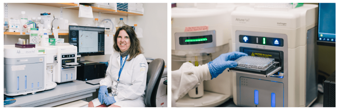
The ThermoFisher Attune (2017 model) is a user-friendly analytical instrument with 4 laser excitation wavelengths 405 nm, 488nm, 561nm, and 635nm. Researchers can measure up to 14 colors simultaneously. This cytometer operates either in a single tube mode or using the autosampler (96 and 384 well, including deep well for around 2 ml/sample). This acoustic focusing-based instrument is highly versatile and can analyze samples with similar sensitivity to other sheath fluid based flow cytometers. In addition it has specially engineered features to analyze 1) very dilute samples 2) larger size particles, and 3) blood constituents without the processing of whole blood. Sample size can vary between 200 ul – 2ml. The Attune software can also be used as analysis software for data analysis of cell populations, statistics, and graphs for documentation and presentations.
The WOLF G2 sorter is a gentle (<2 psi) benchtop microfluidic cell sorting with two lasers and up to nine colors, and has simple workflows for either bulk sorting or single-cell dispensing. Single-cell sorting can be completed in 96- or 384-well plates when using the WOLF G2 in conjunction with its N1 Single-Cell Dispenser. Sorting is done using a self-contained disposable cartridge. The system is ideal for use in many different research fields and application areas like single-cell genomics, cell line development, gene editing, stem cell sorting, cells or organisms requiring specialized media conditions, antibody discovery, immunology, infectious disease, basic research, and more.
Proteomics
The Bio-Plex MAGPIX multiplex reader is a compact, robust system for magnetic bead–based immunoassays. This multiplex reader is capable of reading assays designed on magnetic xMAP (MagPlex) beads. Read up to 50 analytes per sample (as little as 25 ul), and can be used to quantify levels of phosphoproteins and cytokines that are present in cell or tissue lysates, serum or plasma samples. It can also be used to analyze both proteins and nucleic acids. Researchers can quickly evaluate analytes by using pre-validated assays available in the areas of cancer, cardiovascular, cell signaling, cellular metabolism, immunology, metabolism and endocrinology, neuroscience, stem cells, and toxicity. Researchers can quickly evaluate analytes by using pre-validated assays available in the areas of cancer, cardiovascular, cell signaling, cellular metabolism, immunology, metabolism and endocrinology, neuroscience, stem cells, and toxicity. Sensitivity is between 1-10 pg/ml – 100,000 or 3.5 log dynamic range.
Great for a non-wet lab who needs to do accurate Protein/Peptide quantitation. The Direct Detect system provides more accurate results without the pitfalls of colorimetric assays. By measuring amide bonds in protein chains, the IR spectrometry system accurately quantitates an intrinsic component of every protein without relying on amino acid composition, dye binding properties or reduction-oxidation (redox) potential. You can evaluate major components of complex mixtures separable from the Amide I & II region, like lipids and carbohydrates—making accurate quantitation of lysates and membrane preps compared to Bradford or BCA assays.
- More compatible with detergents, reducing agents and other buffer components that interfere with traditional protein quantitation assays, providing more information from your sample
- Preserves precious samples—requires only 2 μL per analysis
- Also quantifies long aliphatic chains in lipids and detergents, so good for purification evaluations
Genomics
The CTAF qPCR System features six independently controlled thermal electric units, providing even, precisely controlled temperatures at all times during the run, including ramping. The programmable thermal gradient feature allows a temperature gradient of up to 24°C across the reaction block at any step in a protocol. Uses low-profile strip tubes or plates.
The optical system, with three filtered LEDs and three filtered photodiodes, collects data from all wells, detecting up to two targets per well. For single-color FAM, EvaGreen, and SYBR Green I Assays, the fast-scan option reads single-channel fluorescence in all 96 wells in just 3 seconds. A separate channel with an LED filter–photodiode combination is dedicated to FRET singleplex experiments.
The T100 thermal cycler is a small thermal cycler offering a comprehensive set of convenient features in a small footprint. This compact thermal cycler features an intuitive touch-screen user interface to make running PCR easy. Thermal gradient technology allows you to quickly optimize your reaction in a single run. With its robust design, the T100 system is a reliable personal thermal cycler that delivers exceptional performance for years. Temperature range 4–100°C, with temperature accuracy ±0.5°C of programmed target, and temperature uniformity ±0.5°C well-to-well within 30 sec of arrival at target temperature.
The Gene Pulser Xcell System is a flexible, modular electroporation system for transfecting every cell type from primary, suspension, and difficult-to-transfect cells, including T cells, to bacteria and fungi. The system is composed of a main unit, two accessory modules, the capacitance extender (CE module) and the pulse controller (PC module), and a ShockPod cuvette chamber.
- Preset protocols — ready-to-use pre optimized protocols for frequently used mammalian and bacterial cell types
- Manual operation — allows for entry and storage of new parameters or editing of all parameters
- Optimization protocols — enables the best conditions to be determined using incremental voltage steps
- Exponential and square waveforms — allows for optimal delivery
Histology Room
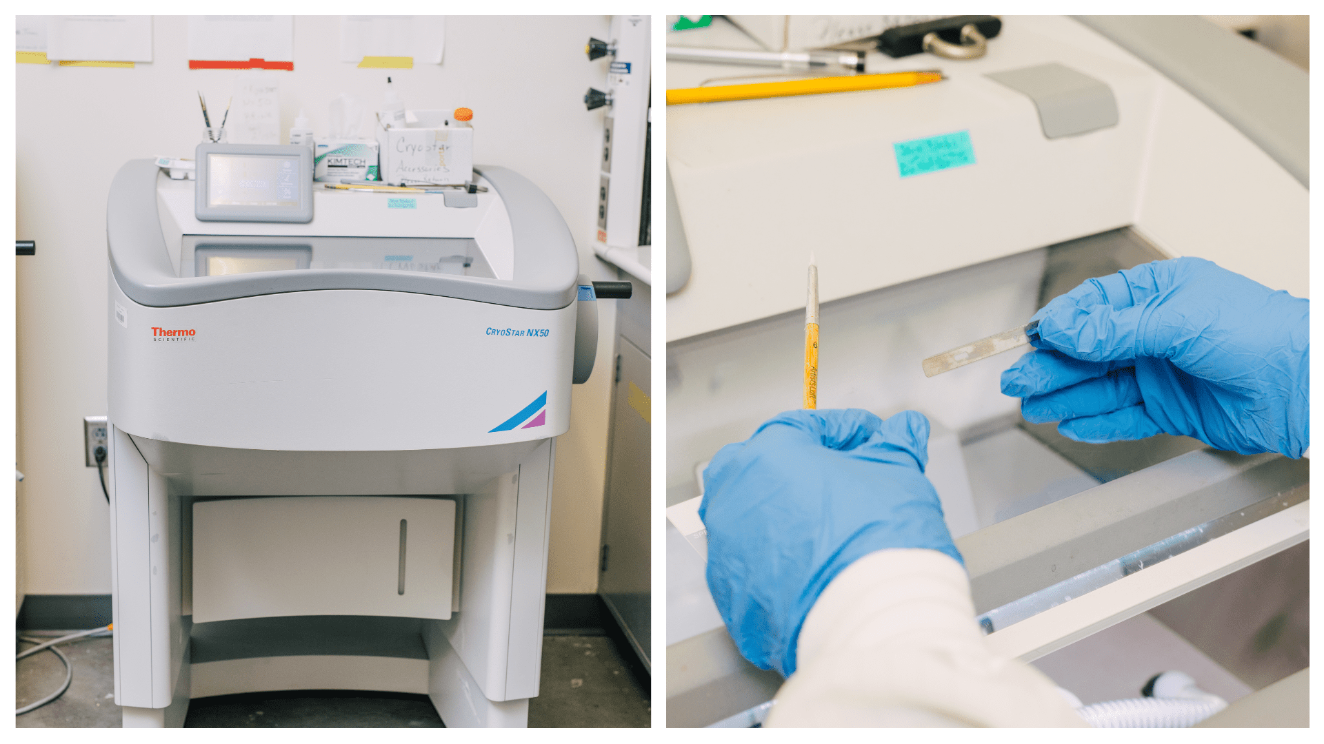
The CryoStar™ NX50 Cryostat is a state-of-the-art cryostat with standard features well-suited to the histology laboratory, including Rapid Response Object Head Temperature Control and a color touch screen user interface for control of instrument functions.
The cryochamber features a spacious stainless steel chamber for workflow organization, chamber cooling to –25 °C, ±2°C (at 20°C ambient), separate specimen head cooling to –43 °C, ±2 °C (at 20 °C ambient), and a continuously cooled knife holder to –27 °C, ±2 °C (at 20 °C ambient). The integrated peltier fast freezing device rapidly cools to –57 °C, ±3 °C. The microtome features a manual light touch rotary flywheel, with sample orientation adjustable 8° in the Y axis for increased stability and 360° Z axis rotation.
The NX50 utilizes a rapid response cooling system that actively cools all areas of the cryochamber that comes in contact with the sample, including the specimen clamp, cryobar, and blade, resulting in precise temperature control. Height adjusts for the cutting chamber so that standing or sitting is comfortable. The CTAF NX50 includes an anti-roll plate, vacutome, and Cold D™ Cold Disinfection which disinfects the cold cryochamber in just 50 minutes.
The ElectroForce® 3200 test instruments was optimized for the characterization of Native Tissues, Scaffolds, and Biomaterials. It features a 225 N maximum force. With the versatility of static to 200 Hz frequency response, the table-top configuration is adaptable to a variety of biomedical research and engineered materials test applications, including torsion testing, creep under dynamic loading and special environments (hot/cold chambers). We have one set of test grippers optimized for our 5 lb load cell, and we also have a 250 g load cell for which accessories may be ordered or expertly machined.