QB3-Berkeley brought the first focused-ion-beam microscope to the UC Berkeley campus in 2011, in collaboration with multiple academic units.
In 2014, a $2 million NSF grant enabled the addition of the first multi-beam helium-ion microscope to be installed at an academic institution in the U.S. The two instruments are housed in QB3’s Biomolecular Nanotechnology Center (BNC) in Stanley Hall.
Ion-beam microscopes are versatile tools used for imaging, analysis, micro- and nanofabrication. In a typical focused ion-beam (FIB) microscope, a gallium-ion beam is finely focused and scanned across the sample for surface imaging and analysis, and at high currents, researchers can perform site-specific sputtering of materials generating features down to the nanoscale. Typically, an electron-beam column is also integrated into the tool for imaging by scanning electron microscopy (SEM), and SEM-based chemical and crystallographic analysis.
Recent advances have lead to the development of a new class of ion-beam microscope known as the helium-ion microscope (HIM), which produces an intense beam of helium ions formed by gas field-ionization at the apex of an atomically sharp tungsten tip. With the HIM, researchers can achieve charged-particle-based surface imaging, analysis and nanofabrication with unprecedented spatial resolution topographic and compositional contrast.
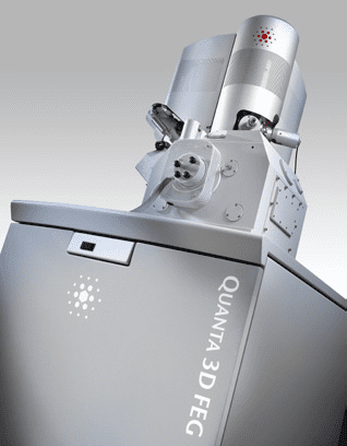 The FIB instrument in the BNC is an FEI Quanta 3D FEG that in addition to the Ga+ beam incorporates a second column for scanning electron microscopy (SEM). Learn more about the dual-beam FIB microscope. The FIB instrument in the BNC is an FEI Quanta 3D FEG that in addition to the Ga+ beam incorporates a second column for scanning electron microscopy (SEM). Learn more about the dual-beam FIB microscope. |
T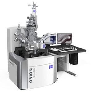 he HIM instrument in the BNC is a Zeiss ORION NanoFab that enables the user to work with He+ or Ne+ beams generated from the HIM source and also incorporates a separate Ga+ FIB column. Learn more about the multi-beam HIM instrument. he HIM instrument in the BNC is a Zeiss ORION NanoFab that enables the user to work with He+ or Ne+ beams generated from the HIM source and also incorporates a separate Ga+ FIB column. Learn more about the multi-beam HIM instrument. |
User Access
Use of the FIB and HIM instruments is available to BNC members. Please visit the BNC website for membership information and rates.